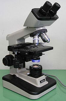
The Microscope and Animal Cells
Homoestatic Imbalance:
One rare disease that can affect muscles during adulthood is myasthenia gravis (mi"as-the'ne-ah gra'vis; asthen = weakness; gravi = heavy), a disease characterized by drooping of the upper eyelids, difficulty in swallowing and taking, and generalized muscles weaknessand fatigability. The disease involves a shortage of acetylcholine receptors at the neuromuscular juncion. The blood of many of these patients contains antibodies to acetylcholine receptors, which suggests that myasthenia gravis is an autoimmune disease. Although the receptors may initially be present in normal numbers, they appear to be destroyed as the disease progresses. Whatever the case, the muscle cells are not stimulated properly and get progressively weaker. Death usually occurs as an result of inability of the respiratory muscle to function. This is called respiratory failure.(for muscular system)
Other Trivias/ Facts:
Since their original invention, microscopes have moved beyond the simple visible light refracting lenses. Electrons, x-rays, and infrared rays are used by far more sophisticated microscopes to detect even smaller and smaller structures. Scanning electron microscopes are able to resolve viruses, which are are smaller than any cell. The cell is the basic unit of life. All organisms are made up of cells (or in some cases, a single cell). Most cells are very small; most are invisible without using a microscope. Cells are covered by a cell membrane and come in many different shapes. The contents of the cell are called the protoplasm. Understanding the study of cells is important because things happen on cellular levels- infections, cancer, transplant rejection, regrowth. In class, We saw different parts of a cell which We found interesting in so many ways. Knowing that if anything abnormal happened to your cells wold be a problem in the human body, not just us human body but also animals and other living things. If We were to want to know more about cells, We'd like to learn more about cells that make plants grow. Other than we have learned a lot in class during laboratory sessions.
If the cerebellum is damaged (for example, by a blow to the head, a tumor, or a stroke), movements become clumsy and disorganized - a condition called ataxia. Victims cannot keep their balance and many appear to be drunk because the loss of muscle coordination. They are no longer able to touch their finger to their nose with eyes closed - a feat that normal individuals accomplish easily. (for nervous system)
Meningitis, an inflammation of the meninges, is a serious threat to the brain because bacterial or viral meningitis may spread to the nervous tissue of the CNS. This condition of brain inflammation is called encephalitis. Meningitis is usually diagnosed by taking sample of cerebro spinal fluid from the subarachnoid space. (for nervous system)
Certain events can cause hair to gray or fall out prematurely. For example, many people have claimed that they turned gray nearly overnight because of some emotional crisis in their life. In addition, we know that anxiety, protein-deficient diets, therapy with certain chemicals (chemotherapy), radiation, excessive vitamin A, and certain fungal diseases (ringworm) can cause both graying and hair loss. However, when the cause of theses is not genetic, hair loss is usually not permanent. (Skin & Body Membranes)